Image of the month

Why you see them, why you don't (Radiographs and uroliths)

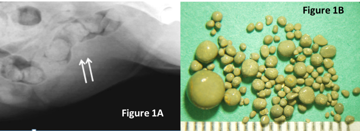
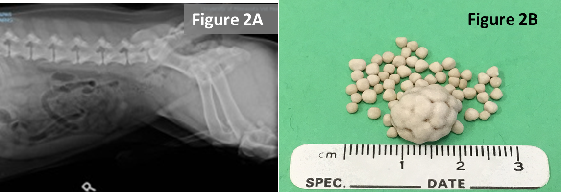
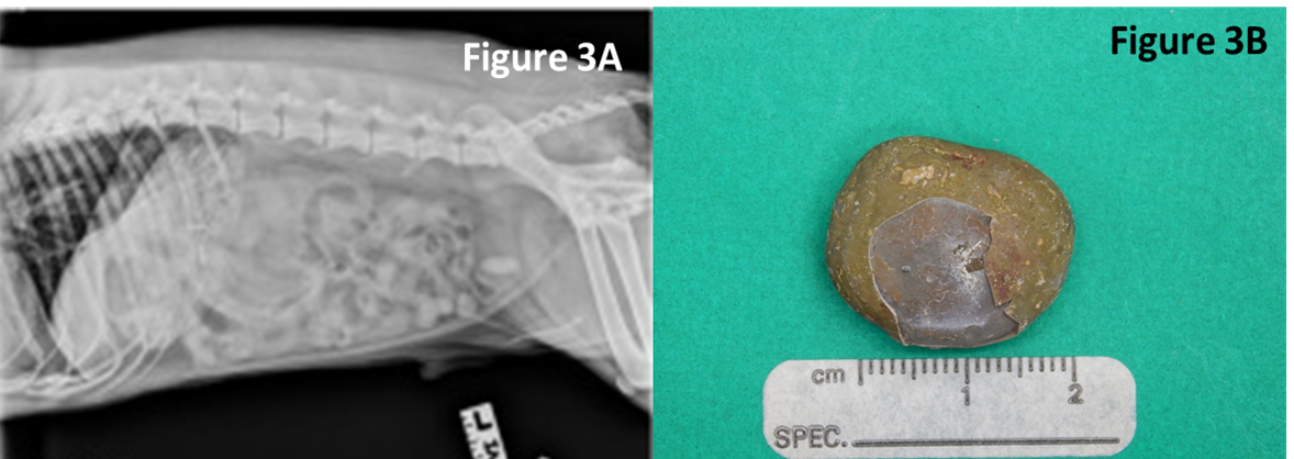
Don’t be fooled by published texts describing urate and cystine uroliths as radiolucent. They are correct that urate and cystine are the least radiopaque of the common stones in dogs and cats. However, radiographic appearance of uroliths depends on several factors of which size and mineral type are the most important. Small urate and cystine stones (Figure 1 and 2) may be radiographically difficult to discern but as they become larger they are more obvious (Figures 2 and 3).  

Things that can be done to improve radiographic discernibility

  1. A full bladder improves contrast
  2. A steady patient improves contrast
  3. Imaging the entire urinary tract avoids missing urethral and kidney stones
  4. Pulling the legs back or forward away from the proximal os penis prevents femurs from obscuring stones
  5. When in doubt additional imaging techniques may be needed (e.g. contrast enhanced radiography, ultrasonography, computerized tomography)
  6. Look at extra-urinary structures (dogs with small livers may be an indication of urate stones in dogs with portovascular shunts.
radiograph of a dog with ammonium urate uroliths and a close up image of the uroliths
Figure 1. Survey radiograph of a female miniature schnauzer with ammonium urate uroliths (A). This close up image of the urinary bladder reveals minimally radiopaque urocystoliths (arrows). Uroliths were removed by voiding urohydropropulsion and consisted of a variety of sizes (B)

radiograph of a dog with cystine uroliths next to the uroliths with a measuring tape to show relative size
Figure 2. Survey radiograph of a male Bassett Hound with cystine uroliths (A). The larger urocystolith is easily discernable.  The smaller uroliths are minimally radiopaque.  There is urethrolith in the os penis. The radiographic technique could be improved pulling the legs caudally away from the proximal os penis. Stones were removed surgically. The largest urolith is approximately 1cm in diameter (B)
radiograph of a dog with a large ammonium urate urocystolith that is obviously radiopaque next to the urolith and a measuring tape showing the approximate diameter of 2 cm
Figure 3.  Survey radiograph of a male mixed breed dog with a large ammonium urate urocystolith that is obviously radiopaque (A). Notice in the cranial abdomen A small liver.  The urolith was removed surgically. The urolith was approximately 2 cm in diameter.
See other images of the month