Image of the month

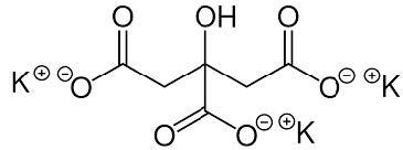
How to adjust potassium citrate to minimize calcium oxalate

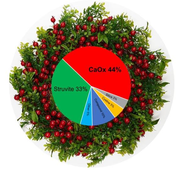
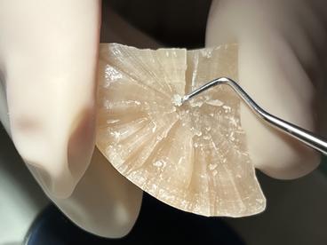
The prevalence of calcium oxalate (CaOx) stones rapidly increased following the implementation of acidifying diets to prevent struvite stones. Potassium citrate, which increases urine citrate and urine pH could reverse this trend. Citrate binds calcium, reducing the amount of calcium available to bind with oxalate.

Citrate reduces CaOx crystal aggregation and aids other crystallization inhibitors. Alkaline urine reduces urine calcium excretion.

Our goal

Potassium citrate is most beneficial in CaOx stone-forming animals with consistently low urine pH ( ≤ 6). Our starting dose is 75mg/kg twice a day to reach a urine pH close to 6.5. Many forms are available. Avoid forms containing cranberries, which are a source of oxalate.

How to adjust the dose

Urine citrate is not routinely measured, but urine pH is. Give pet owners pH paper and have them measure urine pH approximately every 5-6 hours for 2 to 3 days. If the average is near 6.5, no dosage change is needed. For lower values (< 6.0), the dose can be increased by 20% until urine pH approaches 6.5. With adverse effects (decreased appetite, serum potassium > 6.2 mmol/L), the dose is reduced by 20%. We are cautious when administering potassium citrate to animals with dehydration, decreased kidney function, diabetes, and hypoadrenocorticism (disease conditions associated with decreased potassium excretion.

chemical structure potassium citrate
See other images of the month

Image of the month

What species, other than cats, form urethral plugs?

Urethral plugs have been submitted to the Minnesota Urolith Center from many species including cats, dogs, ferrets, raccoons, goats, sheep, and others.

Between 2010 and 2020, 143 urethral plugs from dogs were submitted to the Minnesota Urolith Center. The mineral component of 114 (80%) were struvite. 12 (8%) were composed of miscellaneous material without minerals. Calcium oxalate, calcium phosphate, urate, compound and mixed made up a small number of samples. While plugs were present in a variety of different breeds, 58% were from Pugs.

Clinical evaluation of 9 dogs with plugs (all struvite) at the University of Minnesota, Veterinary Teaching Hospital did not reveal an underlying cause; urinary tract infections were not diagnosed. Like cats, nutritional prevention of struvite was successful in preventing recurrence.  

See our detailed Canine Struvite Risk Management Recommendations

Urethral plug from a 2-year-old castrated male Pug protruding from the distal urethral opening
cystalline portion of the plug was composed of 100% struvite interspersed in a matrix of cellular and proteinaceous material
See other images of the month

Image of the month

What types of dog stones are at the North Pole?

Geography is important when it comes to disease prevalence and management. If you live near a volcano, expect more silica. If you live in Australia, expect more struvite. If you live in Europe, expect more cystine. And if you are helping Santa (i.e. Saint Nikolaas, Kris Kringle, and many other names), we got you covered (10 years of dog stones from North Pole (Alaska)).

pie chart of dog stones from north pole AK; CaOx 44%, Struvite 33%, compound 10%, cystine 5%, silica 3%, other 5%
See other images of the month

Image of the month

Should you culture stones?

gloved hand holding urolith and a metal probe digging into the urolith's center

Stones can form because of a urinary tract infection (e.g. struvite, calcium phosphate carbonate, and ammonium urate), serve as a risk factor for urinary tract infection (UTI), and harbor bacteria that contribute to recurrent UTI. Even though these two diseases (UTI and stones) are closely related, we rarely recommend stone culture. Culturing the urine before antibiotic therapy is your best tool for assessing the patient and the disease.

Below are guidelines that may help you decide if want to culture the stone when pretreatment urine cultures were not obtained.

  1. If all stones were removed, there is no need to culture the stone. Even if stones were culture positive, we do not recommend treating the source when it has been eliminated. Culture urine 7 to 14 days after ending postoperative antibiotics (or sooner with persistent UTI signs) to assess the need for subsequent antibiotic prescriptions.
  2. If you want to prioritize dietary therapy or UTI control as your principal method to prevent struvite/calcium phosphate carbonate uroliths, culture the stone. Disease epidemiology can also help you decide.  Infection-induced struvite is very common in dogs, especially females; prioritize UTI control in these cases. Sterile struvite is very common in cats, prioritize nutritional prevention in these cases. Still unsure, do both (UTI control and nutritional prevention).
  3. If all stones were not removed, peri- and postoperative antibiotics may change bacterial susceptibility in the patient and the stone. Culture urine 7 to 14 days after ending postoperative antibiotics (or sooner with persistent UTI signs) to assess the need for subsequent antibiotic prescriptions.

In almost all cases, culturing the urine before starting antibiotics & removing the stone is better than culturing the stone. It allows you to select effective antibiotics at the beginning of treatment.

See other images of the month

Image of the month

Are xanthine stones dissolvable?

The answer depends on the underlying cause. Xanthine, an intermediate-byproduct of purine catabolism, is rarely found in urine or stones because it is metabolized to uric acid and then allantoin, a water soluble end-product that is excreted in urine. Hereditary Xanthinuria is a rare autosomal recessive disease caused by loss of xanthine dehydrogenase (XDH) or molybdenum cofactor sulfurase (MOCOS) genes. Acquired Xanthinuria is a potential adverse consequence of allopurinol, a xanthine dehydrogenase inhibitor administered to treat leishmaniasis and prevent urate uroliths. In acquired xanthinuria, xanthine uroliths are dissolvable. Hereditary xanthine uroliths are not dissolvable.

the head of a dalmatian

Case Example: Jake is a 2-year-old male Dalmatian diagnosed with urate uroliths. Following urolith removal, Jake was prescribed allopurinol (15mg/kg /day) to reduce urate stone recurrence. However, a diet change was not recommended. A 2-month recheck identified urolith recurrence (figure 1); uroliths were much larger than his previous ones. During the recheck, small uroliths were retrieved with a urinary catheter and submitted for quantitative analysis. Recurrent stones were composed of 100% xanthine.  With this new information, allopurinol was discontinued and Jake was prescribed a low purine, urine-alkalinizing diet (Prescription diet u/d). Radiographs taken one month later revealed that all xanthine stones had dissolved (figure 2). To prevent future urate urolith recurrence, allopurinol was prescribed at a lower dose (7 mg/kg/day) and the low-purine diet was continued. Read more on allopurinol and urate uroliths (PDF).

double contrast cystogram showing xanthine uroliths in a dog's bladder
Figure 1

Figure 1. Double contrast cystogram showing xanthine uroliths in the urinary bladder of a male Dalmatian following allupurinol administration.


double contrast cystogram showing no uroliths in a dog's bladder
Figure 2

Figure 2. Double contrast cystogram of the dog in figure 1 one month after stopping allopurinol and feeding a low purine diet. Xanthine uroliths rapidly dissolved.

See also our Canine xanthine recommendations (PDF)

See other images of the month

Image of the month

How long should struvite-prevention diets be fed to cats?

close-up of two feline struvite stones on a green background with a ruler underneath showing each stone is over 1 cm long
Figure 1

Nutritional dissolution and prevention of struvite (figure 1) is an innovative compassionate therapy that is rapidly replacing expensive painful surgical removal of struvite stones. Requiring only a few weeks to dissolve struvite stones, how long should therapeutic foods be fed to prevent their recurrence? It will likely vary with each cat, but data from the Minnesota Urolith Center indicates that struvite stones primarily affect young to early-middle age cats (figure 2).  In 2021 we analyzed stones from almost 20,000 cats.  Approximately half were composed of struvite, and of that half, 90% were from cats 10-years-old or younger.  This indicates that for many struvite stone formers, prevention is less needed as cats age.  After switching to a different diet, monitor the cat’s urine a month later. If struvite crystalluria persists, the struvite-prevention diet may need to be fed longer.


Bar graph showing a bell curve of the prevalence of struvite in cats by age in years, with the prevalence highest between ages two and six
Figure 2

See also our Feline Struvite Dissolution  and Feline Struvite Risk Management recommendations 

See other images of the month

Image of the month

What is the significance of urate in canine struvite stones?

a yorkie dog and compound uroliths. Urate nidus with a struvite body

The chemical composition of a urolith is a biomarker for underlying disease. A urolith composed of two calculogenic salts is an indication that two diseases are occurring simultaneously. Dogs with a struvite-urate urolith are often diagnosed with a urinary tract infection and hyperuricosuria (increased urate excretion).  Most infections are caused by Staphylococcal that break down urea to ammonia.  Hyperuricosuria is often caused by genetic variants in a urate transporter or hepatic insufficiently due to vascular anomalies.  It is unknown if all struvite-urate stone formers fit this disease phenotype. To identify those that do, we recommend bile acid testing and genetic testing for urate transport anomalies.

How to manage these two diseases and reduce stone recurrence

  1. Promptly recognize and eradicate urinary tract infection.
    1. Eliminate risk factors (peri-vulvar dermatitis, immunosuppressive drugs, obesity, others).
    2. Teach owners to recognize signs of infection and to seek care early.
    3. Periodic urine cultures (1 month after stone removal and then every 3 to 6 months as needed).
  2. Determine if hyperuricosuria results from a liver or genetic disease.
    1. Perform serum bile acids in dogs with suspected hepatic insufficiency (Yorkshire terrier, miniature Schnauzer, Maltese, pug, shih tzu).  If positive, consider nutritional, pharmacological and surgical strategies depending on disease severity.
    2. Submit a genetic tests for Hereditary Hyperuricosuria (vgl.ucdavis.edu/test/hyperuricosuria) in suspected breeds (e.g. bulldog, black Russian terrier, Labrador, American Staffordshire terrier, Weimaraner, others).  Affected dogs may benefit from low dose allopurinol (5mg/kg daily) especially with recurrent struvite-urate stones.
  3. Feed low purine foods. Since most of these stones are predominantly struvite, it is tempting to feed struvitolytic diets and ignore the urate.  Additional research is needed to determine if this is advantageous, especially when urate is less than 10-20% of the stone.  Until then, we recommend avoiding struvitolytic diets because the urine acidification that these diets produce, promotes urate stone formation.  Instead, we recommend feeding low purine diets (Derm Complete, u/d, HA Hydrolyzed, others), or liver support foods if needed (L/d, g/d, Hepatic).
See other images of the month

Image of the month

What bacteria cause infection-induced struvite stones, which are common in dogs?

Of the approximately 65,000 canine stones analyzed by the Minnesota Urolith Center in 2021, 41% were struvite of which most were infection-induced. In female dogs, struvite is the most common urolith type, representing 74% of submissions. The formation of infection-induced struvite stones depends on the generation of both ammonium ions and alkaline urine. These conditions are met in clinical cases through the action of urease-producing bacteria. The most common bacteria isolated from the urine of dogs with struvite stones or cultured from struvite stones was Staphylococcus spp., which occurred in more than 85% of evaluations (1-3). Many Staphylococcal bacteria produce urease, the enzyme that hydrolyzes urea into ammonia. Ammonia subsequently reacts with water producing ammonium ions and hydroxide ions, the latter of which alkalinizes the urine (see the making of infection-induced struvite below). In humans, Proteus spp. is the most common urease-producing bacterium causing infection induced struvite stones.

The making of infection-induced struvite uroliths

Step 1: Urea (NH2CONH2) +H2O Urease 2NH3 +CO2
Step 2: NH3 + H2O→NH4 +1 + OH-1 (OH -1 producing alkaline urine)
Step 3: H2PO4 -1 + OH-1 → HPO4 -2 + OH-1 → PO4 -3
Step 4: Increasing urine concentrations of [NH4 +1] and [PO4 -3] with [Mg] favors formation of struvite (MgNH4PO4•6H2O)

To dissolve infection-induced struvite, administer appropriate antibiotics to eradicate infection (based on urine culture and sensitivity) and provide therapeutic foods to acidify the urine and lower phosphorus, magnesium, and urea until uroliths are no longer detected on imaging. The average time to complete dissolution of struvite uroliths in dogs is approximately 5 weeks (2,3).

Canine Struvite Dissolution
Canine Struvite Risk Management

Perry et al. J Vet Diagn Invest. 25;2013:199-202.
Dear et al. BMC Vet Res. 2019;15:273.
Wingert et al. J Vet Intern Med. 35;2021;2287-2295.

See other images of the month

Image of the month

How common are canine kidney stones?

In 2021 the Minnesota Urolith Center analyzed stones from over 64,000 dogs. Only 0.3% were from the kidney or ureter. Of these, 36% were calcium oxalate, 27% were struvite, 13% were urate, 5% were cystine, 2% were calcium phosphate, and 5% were composed of multiple minerals.

Does this mean that kidney stones are not common? Likely not. A recent study at the University of Minnesota revealed that 41% of dogs with lower urinary tract urolithiasis had radiographic findings consistent with upper urinary tract stones.1  In a subset analysis of 5 small breeds at risk for CaOx (Bichon, Pomeranian, Lhasa, Shih Tzu and Miniature Schnauzer), 81% of dogs with lower urinary tract urolithiasis had radiographic findings consistent with upper urinary tract stones.

Why are so few kidney stones submitted for analysis? The majority of canine kidney stones do not appear to cause clinically significant disease requiring their removal and subsequent analysis. The ACVIM urolith consensus advices that “only those nephroliths contributing to outflow obstruction, recurrent infection, pain, and those enlarging to the point of causing renal parenchymal compression, should be considered for removal.”2 Uroliths amenable to medical dissolution (e.g. struvite) should be dissolved.

Our clinical advice: When stones are detected in the urinary bladder, remember that all portions of the urinary tract are susceptible and should be included in your imaging protocol to provide comprehensive care and appropriate monitoring.

survey radiograph of a 9-year-old female-spayed dog with multiple struvite stones in the urinary bladder and a stone in the left kidney (arrow)
  1. Hoelmer et. al. Vet Sci 2022;9:283 
  2. Lulich et. al. J Vet Intern Med 2016;30:1564 
See other images of the month
Junshu Yang

Junshu Yang

Researcher 3