Image of the month

Spooky urolith centers

This month we are focusing on the interesting, strange and scary things we have found in the center of uroliths.

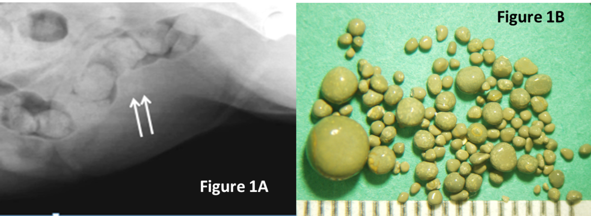
  • Suture was identified in approximately 10% of recurrent stones. When closing the bladder after a cystotomy, minimize placing suture in the bladder lumen to reduce its contribution to stone recurrence.  Image A 
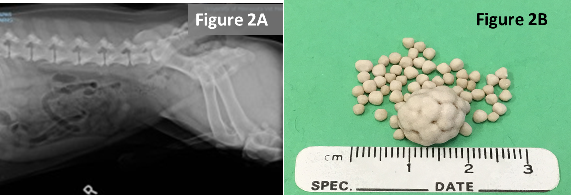
  • Plant awns/material are common in the western United States and sometimes enter the bladder and can become encrusted with mineral.  Image B
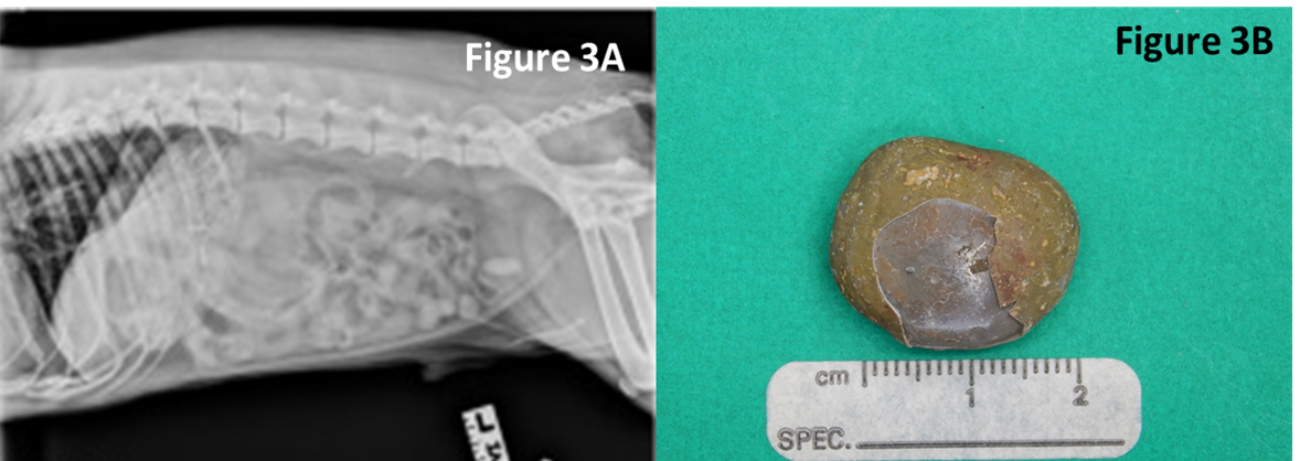
  • Toothpick - a migrating toothpick could have been swallowed by the pet because it held a tasty Halloween treat.  In this case, the toothpick pierced the intestine, traveled to the bladder and initiated stone formation.  Image C
  • Fleas, louse, and other insects may climb up the urethra and become entombed in stones.  Image D
4 uroliths with different foreign body nidus, referenced above as image a-d

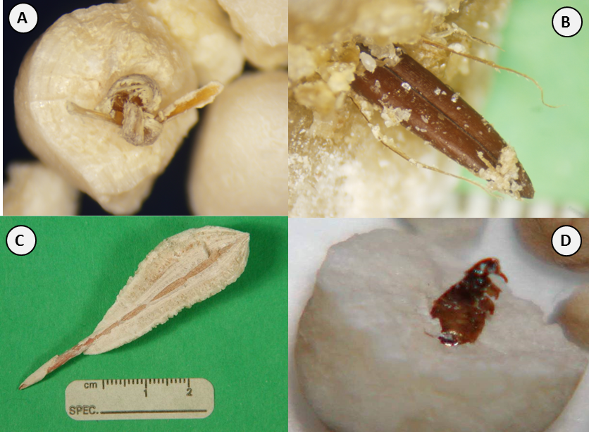
What do all of these spooky invaders have in common?  

Heterogeneous nucleation: the process where crystallization is enhanced by initiating over foreign-preformed surfaces.

What’s in the center of your uroliths?  

For complete and accurate urolith analysis – please submit the entire urolith.

See other images of the month

Image of the month

Feline urethral plugs

Feline urethral plugs are a common cause for urethral obstruction in the male cat. Typically, plugs contain mineral and organic matrix (e.g. inflammatory reactants, blood, sloughed tissue). Understanding their epidemiology and composition are important to prevent their formation and minimize their recurrence.

Between 2011 and 2018 the Minnesota Urolith Center received over 2000 urethral plugs for quantitative analysis. 82% were retrieved from cats younger than 9-year-old. Varieties of minerals were identified in urethral plugs. 92% were struvite. This information is critical to manage urethral obstruction in cats.

Consider these points.

  • Submit plugs for mineral analysis to identify risks contributing to urethral obstruction and ways to prevent future obstructions.
  • Almost all plugs were composed of struvite.  To prevent recurrence, feed struvite dissolution foods.
  • Feed struvite dissolution foods until cats are approximately 9 to 10-years-old. When stopping therapeutic foods, repeat urinalyses before and several months after food change. Recurring struvite crystals is an indication that struvite dissolution foods should be fed longer.
  • To avert first-time, life-threatening urethral obstructions, feed struvite dissolution foods in young male cats, especially those with persistent struvite crystalluria.
Figures 1 through 4 referenced below

Figure 1. Lateral radiograph of a 2-year-old cat presenting for inability to urinate. Accumulation of radiodense sand within the pelvic to penile urethra (arrows) identified the cause of the obstruction as a urethral plug.

Figure 2.  Urethral plug passed by the cat in figure 1. The mineral component was 100% struvite. Each tick mark equals 1 mm.

Figure 3.  Lateral radiograph of the cat in figure 1 two weeks after passing his urethral plug and consuming Hills Prescription food c/d multicare.  No mineral densities are observed in the urinary bladder or urethra.

Figure 4.  Age distribution of cats whose urethral plugs were submitted to the Minnesota Urolith Center between 2011 and 2018 for quantitative mineral analysis.

See other images of the month

Image of the month

Urolith basics

What are uroliths?

Uroliths (also known as calculi or stones) are solidified aggregates of mineral and nonmineral crystalloids that form in the urinary tract when urine becomes oversaturated with crystallogenic precursors. Most uroliths are composed of biogenic minerals, but also form from drug metabolites, amino acids or over foreign substances such as suture material from a previous surgery.

uroliths of different composition from dogs and cats next to a struvite stone with a suture nidus

What is the anatomy of a urolith?

diagram of the structures within a urolith, nidus, body, shell, and surface

Structure

Description Significance
Nidus Area of obvious initiation of urolith growth, which is not necessarily the geometric center of the sample.

In general, the mineral type(s) in the "nidus" layer should be the primary focus of preventative measures because the nidus is the area of urolith initiation. Preventing a nidus of foreign material such as a suture, plant, etc. is also important to prevent urolith recurrence.

Body The major portion of the urolith. The "body" layer comprises the largest bulk of the sample. If no nidus layer is listed (or the nidus is similar in composition to the body layer), this should be the focus of preventative measures.
Shell A complete outer concentric lamination of the urolith. Shell and/or surface layers represent the most recent activity in the urolith formation process. Often, shell and surface layers that are significantly different from the composition of the body layer reflect changes in diet, medication, or the patient's medical condition.
Surface An incomplete outer lamination of the urolith. See "Shell" significance above.


Why submit uroliths for analysis?

Survey radiograph of a dog with a urate urolith and a small liver consistent with a vascular shunt of the liver

Developing strategies to minimize urolith recurrence requires knowledge of its composition. Urolith composition is a clue to the underlying diseases responsible for their formation. Submit all or a representative number of uroliths to obtain an accurate analysis. How to submit uroliths

What methods of analysis are recommended?

  1. Quantitative analytical methods are most accurate. The Minnesota Urolith Center uses optical crystallography, infrared spectroscopy, x-ray diffraction, energy dispersive techniques, and other methods to quantitatively analyze each layer in uroliths.
  2. Qualitative analytical methods are less reliable.  This method uses chemical tests to identify chemical radicals and ions. Several components in stones cannot be identified chemically.

How can uroliths be retrieved for analysis?

  • Voluntary voiding
  • Catheter retrieval
  • Voiding urohydropropulsion
  • Basket retrieval
  • Laser lithotripsy and basket retrieval
  • Percutaneous cystolithotomy/nephrolithotomy and basket retrieval
  • Surgery

For more information about retrieval techniques visit our publication page under urolith retrieval methods.

What steps should be taken to minimize urolith recurrence?

The Minnesota Urolith Center provides detailed diagnostic and therapeutic strategies to assist veterinarians and pet parents in caring for dogs and cats with uroliths and other urinary disorders. See our Treatment Recommendations

See other images of the month

Image of the month

Recognizing urinary tract disease in the time of COVID

Taking a pet to the veterinarian during a pandemic has created new challenges.

Is seeing a veterinarian necessary? Can I wait? What is the best way to communicate my concerns? Here are several tips to make this pandemic easier to manage for pets even after the pandemic has ceased.

Some supplies you will need

  • A smart phone: Take a picture or movie of your pet urinating. Include the size and location of the urine accident, and the color of the urine.
  • A soup ladle: This is a great tool to catch a midstream urine sample in dogs. Walking dogs outside the veterinary clinic while waiting to get inside may induce your dog to urinate. Urine is an important sample to evaluate. Assist your veterinarian by bringing in a clean urine sample. Collect it when you leave the house. Store it in a clean container with a lid.
  • Urine dipsticks: Urine test strips are inexpensive, purchased online, and can detect blood in 60 seconds. Blood is one of the most common signs of urinary tract disease, and an indication that your pet should see your veterinarian sooner than later.
  • Motion activated camera: in multiple cat households, it is difficult to see who is urinating inappropriately. Motion activated cameras can catch the culprit in the act so that you know which pet to bring in for care.

See a 20 minute webinar for more information

See other images of the month

Image of the month

Does water type matter?

Why add water?

Increasing dietary water from 7% to 73% decreased calcium oxalate relative supersaturation (a surrogate marker for stone formation) in normal dogs and cats (Stevenson 2003 & Buckley 2011).

Is hard water detrimental?

map of the United States with color-coding to show that the eastern seaboard has the least CaCO3, while the western seaboard has the most CaCO3has
Figure 1

The answer may be surprising. Water hardness is determined by its quantity of calcium carbonate. Soft water is <60mg/L and very hard water is >180mg/dl. Tap water varies in mineral and electrolyte content between geographic areas even in the same country (Figure1).  In human stone formers, hard water increased calcium excretion, but either resulted in no difference or a decrease in rates of stone formation (Sulaiman 2020). Other factors such as magnesium and bicarbonate content may be responsible for this paradoxical finding (i.e. higher urine calcium associated with lower calcium stone rates).

Clinical impact

Additional water intake has a beneficial effect in preventing calcium oxalate stones (add 1 cup of water to every cup of dry canine c/d multicare to achieve a moisture content of >73% or fed c/d multicare canned which is >77% moisture). Paradoxically, hard or tap water with higher calcium, magnesium and bicarbonate or other alkaline buffers may be preferable to distilled water or soft mineral water. Studies in dogs and cats with urolithiasis have not been performed.

References

See other images of the month

Image of the month

Why you see them, why you don't (Radiographs and uroliths)

Don’t be fooled by published texts describing urate and cystine uroliths as radiolucent. They are correct that urate and cystine are the least radiopaque of the common stones in dogs and cats. However, radiographic appearance of uroliths depends on several factors of which size and mineral type are the most important. Small urate and cystine stones (Figure 1 and 2) may be radiographically difficult to discern but as they become larger they are more obvious (Figures 2 and 3).  

Things that can be done to improve radiographic discernibility

  1. A full bladder improves contrast
  2. A steady patient improves contrast
  3. Imaging the entire urinary tract avoids missing urethral and kidney stones
  4. Pulling the legs back or forward away from the proximal os penis prevents femurs from obscuring stones
  5. When in doubt additional imaging techniques may be needed (e.g. contrast enhanced radiography, ultrasonography, computerized tomography)
  6. Look at extra-urinary structures (dogs with small livers may be an indication of urate stones in dogs with portovascular shunts.
radiograph of a dog with ammonium urate uroliths and a close up image of the uroliths
Figure 1. Survey radiograph of a female miniature schnauzer with ammonium urate uroliths (A). This close up image of the urinary bladder reveals minimally radiopaque urocystoliths (arrows). Uroliths were removed by voiding urohydropropulsion and consisted of a variety of sizes (B)

radiograph of a dog with cystine uroliths next to the uroliths with a measuring tape to show relative size
Figure 2. Survey radiograph of a male Bassett Hound with cystine uroliths (A). The larger urocystolith is easily discernable.  The smaller uroliths are minimally radiopaque.  There is urethrolith in the os penis. The radiographic technique could be improved pulling the legs caudally away from the proximal os penis. Stones were removed surgically. The largest urolith is approximately 1cm in diameter (B)
radiograph of a dog with a large ammonium urate urocystolith that is obviously radiopaque next to the urolith and a measuring tape showing the approximate diameter of 2 cm
Figure 3.  Survey radiograph of a male mixed breed dog with a large ammonium urate urocystolith that is obviously radiopaque (A). Notice in the cranial abdomen A small liver.  The urolith was removed surgically. The urolith was approximately 2 cm in diameter.
See other images of the month

Image of the month

1.5 million stones and counting

our 1.5 millionth urolith, patient and radiograph

The Minnesota Urolith Center just analyzed its 1.5 millionth stone. The stone was submitted by the Savanna Animal Hospital in Jensen Beach, Florida. Brody, an 11-year-old, male-neutered, miniature Schnauzer appeared uncomfortable and shaking. An X-ray of the abdomen quickly identified the cause. The calcium oxalate stone was removed and Brody’s parents say that he is like a puppy again.

This year, the Minnesota Urolith Center celebrates its 40th anniversary. Each year we analyze 90,000 samples from around the world. We adopted a Wikipedia business model before Wikipedia became famous. That is, we rely on donations from individuals, clinics and corporations to provide our service at no cost to veterinarians and the pets they serve. In 2007, during the great recession, donations dropped to almost 50%; we wondered if we had to close our doors. With time, increasing donations, and a large educational gift from Hills Pet Nutrition, we never stopped supporting you.  We are excited to be a part of the veterinary profession because we are driven by your compassion to give.

Minnesota Urolith Center - celebrating 40 years of service 1981 - 2021
See other images of the month

Image of the month

Cystine increasing

Canine cystine is on the rise. In 2000, cystine uroliths were uncommon (0.8 %). In 2020, the number of cystine submissions octupled (6.8%)!

bar chart of the percent of canine cystine submissions by 5 year increments between 2000 and 2020 showing an large increase in submissions between 2015 and 2020
multiple cystine uroliths


Cystine is an amino acid that is filtered in urine and reabsorbed by the renal tubules (i.e. removed cystine from the urine). Cystine uroliths result from high urine cystine levels due to cystine transportation defects in renal tubules. The defect has been identified in several genes (autosomal recessive-SLC3A1 in Labradors, Newfoundlands, and Landseers, autosomal dominant-SLC3A1 in Australian Cattle dogs, autosomal dominant-SLC7A9 in the Miniature Pinschers, and sex linked/androgen responsive in Mastiffs, English and French Bulldogs). These breeds can be genetically tested (research.vet.upenn.edu/penngen). Many breeds without an available genetic test have androgen-responsive cystinuria. To determine if dogs with cystine uroliths have androgen-responsive cystinuria, measure urine cystine (UC Davis Amino Acid Laboratory) and urine creatinine (at your routine lab) before and three months after castration. In androgen-responsive dogs, the cystine to creatinine ratio drops significantly at the three-month recheck.

Learn more about cystine (Canine Cystine Uroliths and supplemental information and Why you see them, why you don't (about radiographs and uroliths))

See also Step by Step: Risk Management for Canine Cystine Uroliths Image of the Month, Feb 2023

See other images of the month

Image of the month

How to calculate urine cystine:creatinine ratio?

Androgen-dependent cystinuria is a common cause for cystine uroliths in dogs that is effectively treated with castration. Genetic testing is available for Mastiffs, English Bulldogs and French Bulldogs. However, many other breeds without an available genetic test may also have androgen-dependent cystinuria. To identify these dogs, we compare urine cystine:creatinine ratios before castration and 3 to 6 months after castration. Figure 1 shows the changes in urine cystine:creatinine ratios in a castrated dog without androgen-dependent cystinuria and recurrent cystine uroliths (orange line that remains elevated) and an androgen-dependent cystinuric dog immediately before (time = 0) and after castration that never reformed cystine uroliths (blue line showing a gradual reduction.

line graph showing the urine cystine (nmol/mg of creatinine) in a castrated dog without androgen-dependent cystinuria (orange line that remains elevated) and an androgen-dependent cystinuric dog, before and after castration (blue line showing a gradual reduction)

A spot (random) urine cystine:creatinine ratio is a simple alternative to measuring 24-hour urine cystine excretion. To calculate this ratio the denominators in the concentrations of cystine and creatinine must be identical. If cystine is reported in nmol/ml and creatinine in mg/dl, convert the creatinine concentration to mg/ml by dividing the creatinine by 100 (i.e. one dl = 100 mls). Then divide the cystine by the converted creatinine concentration. For example, in a dog with a urine cystine (measured at UC Davis Amino Acid Laboratory) of 389 nmol/ml and a urine creatinine (measured at internal laboratory) of 206 mg/dl convert the creatinine to mg/ml (=206/100 =2.06). Divide the cystine (389 nmol/ml) by the converted creatinine (2.01mg/ml) resulting in a cystine:creatinine ratio of 188.8 nmol of cystine/mg of creatinine.

Learn more about canine cystine:

See other images of the month

Image of the month

Step by step: Risk management for calcium oxalate (CaOx)

a calcium urolith image over a radiograph of a dog with a calcium oxalate urolith
  1. Correct idiopathic hypercalcemia.  Although rare in dogs, it is more common in cats (30%).  We do not recommend bisphosphonates or calcimimetics to lower idiopathic hypercalcemia when uroliths are the only reason. The side-effects of these medications can be worse than the uroliths. Consider a diet change where dietary calcium is less than 200 mg/100kcal and the Ca:P ratio is less than 1.4:1 (Ehrlich 2024).  If ineffective, add 2 grams of Chia seeds to the cat’s food daily (Fantinati 2020).  Some cats revert without therapy.
  2. Select a CaOx prevention food. Pet food manufacturers meticulously tailor ingredients and test their foods to ensure a reduced urine CaOx precipitation while providing good nutrition.
  3. Reduce urine specific gravity. In healthy dogs, decreasing urine specific gravity from 1.034 to 1.024 reduced CaOx saturation by 34% (Stevenson 2003). In healthy cats, decreasing USG from 1.053 to 1.036 reduced CaOx saturation by 50% (Buckley 2011). We recommend feeding canned foods or adding water to food to achieve a urine specific gravity below 1.020 in dogs and 1.030 in cats.
  4. Neutralize urine pH.  In healthy cats, alkaline urine (pH=7.03) reduced CaOx saturation by 79% (Bartges 2013). Dogs with aciduria were at higher risk (OR = 1.94) for their first CaOx stone (Okafor 2014). We recommend administering potassium citrate to achieve a urine pH between 6.5 to 7.5. 
  5. Medically image every 6 to 12 months.  CaOx is a common and difficult stone to prevent because factors responsible for formation are not completely understood. Stone recurrence detected early, prior to clinical signs and when uroliths are small enough to pass through the urethra can be removed non-surgically by voiding urohydropropulsion.
  6. Administer hydrochlorothiazide diuretics with highly recurrent stones (recurrence in less than 1 year).  Hydrochlorothiazides reduce urine calcium excretion.

See our detailed Canine Calcium Oxalate and Feline Calcium Oxalate Risk management recommendations.

See also:  How to adjust potassium citrate to minimize calcium oxalate

See other images of the month