A roundup on radiomics: the machine learning technique shows promise for diagnosis of canine tumors
October 18, 2021

Editor's note: Radiomics is a relatively new method of extracting potentially unseen data from medical imaging techniques. In this method, machine-learning algorithms assess diagnostic images from tools such as CT and arrive at features that might add up to, say, a specific tumor. Put another way, radiomics is a quantitative description of a particular image that spells a noninvasive diagnostic and treatment strategy.
It appears no field—least of all medicine—is spared from the increasing need to digitize and analyze greater and greater amounts of data, and radiomics is one of the new, sophisticated tools that allows medical professionals to do so. Though radiomics has shown good promise in diagnosing human masses, little has been published on radiomics-related studies of canine lung and liver tumors. Below is a glimpse into a study indicating the method holds promise for animal health, much as it appears to for humans. Next week, we'll look at how radiomics can help detect liver masses in dogs.
Research roundup: Radiomics offers new ways of looking at tumors to guide clinicians treating lung cancer
The most commonly diagnosed and most deadly cancer in humans worldwide is lung cancer. Current lung cancer treatments often involve custom therapies tailored to a tumor’s specific cell type and characteristics. Identification of the tumor's physical and cellular profile so that a diagnosis, treatment plan, and prognosis can be provided usually requires a tissue sample obtained via a costly and invasive lung biopsy.
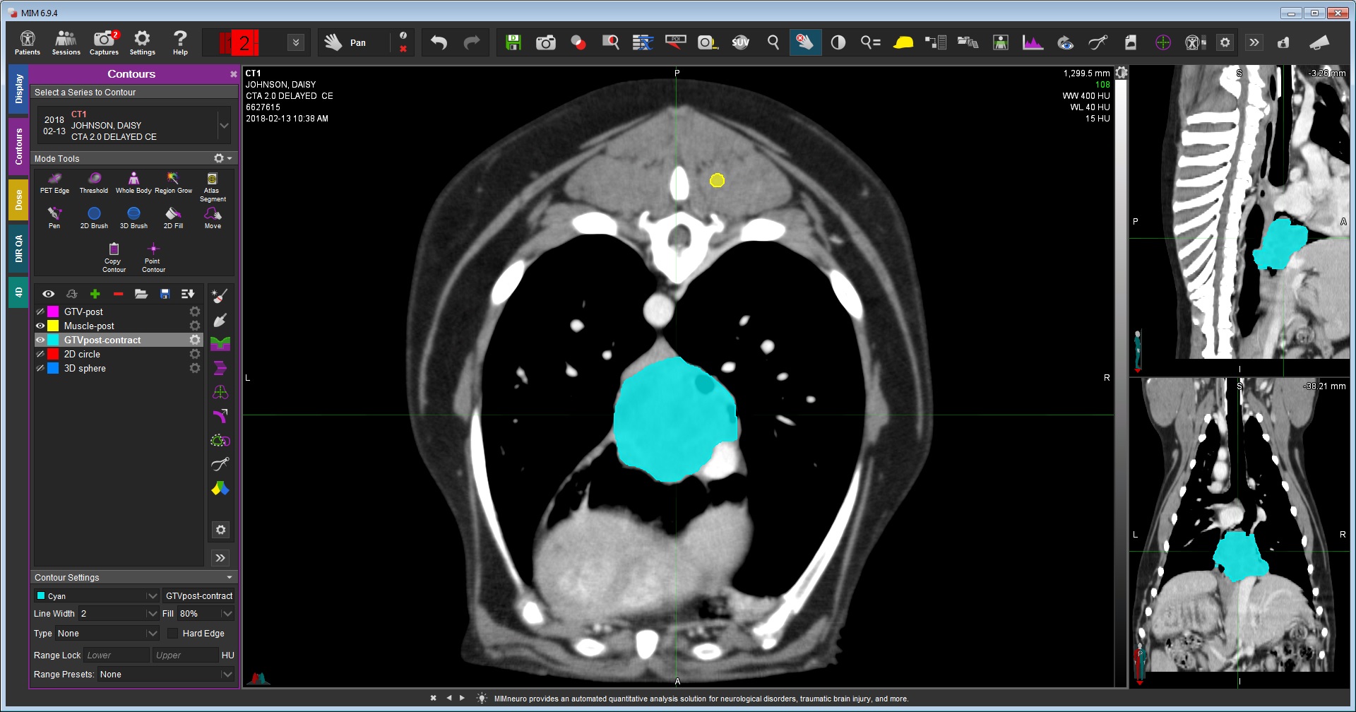
Seeking an objective, noninvasive, and reliable method for diagnosis and treatment, a team of researchers at the University of Minnesota’s College of Veterinary Medicine (CVM) including Jessica Lawrence, DVM, DACVIM (Onc), DACVR (Rad Onc), DECVDI (add on Rad Onc), Amber Wolf-Ringwall, DVM, PhD, and Hannah Able, DVM, conducted a first-of-its-kind study to assess CT radiomic features in dogs with lung cancer. Radiomics is a relatively new area of medicine that extracts measurable data from CT images of the tumor and applies statistical analysis to uncover patterns and characteristics not always detected by the naked eye. The method can predict a tumor’s physical characteristics like shape and volume, as well as the cellular makeup, without a lung biopsy.
Mining electronic medical records, CT images, and tissue samples collected from 65 dogs with a primary lung tumor diagnosis over a 10-year period at the CVM’s Veterinary Medical Center, the researchers used radiomics to collect additional tumor data from the original CT images. The data included tumor diameter, volume, shape and texture, and more. The physical tumor characteristics identified by radiomic data were then considered in light of the original diagnoses, prognoses, and outcomes established via traditional biopsy, pathology, and radiology techniques. The study found radiomic analysis could accurately differentiate between some cancer types such as adenocarcinoma and non-adenocarcinoma. Because different cancers have varying treatment needs and survival times, using radiomics as a diagnostic tool can inform clinical practice before a biopsy or surgical removal, and it may help dogs that would benefit from additional diagnostics or certain treatment types.
Certain tumor characteristics collected such as volume, diameter, and intensity correlated with prognosis and outcomes reflected in the patient’s original clinical records. These features, when combined with other clinical information, can be correlated with clinical-outcomes data previously noted and used by clinicians to make evidence-based decisions going forward.
Radiomics identifies tumor characteristics on imaging that could potentially aid cancer detection, diagnosis, assessment of prognosis, prediction of response to treatment, and monitoring of disease status. Future radiomic studies will require standardized workflows and protocols to obtain best results. And radiomic features should continue to be evaluated and their workflows optimized in future canine clinical trials to better refine the prognostic use of radiomics in cancer care.
Since canine and human diagnostic and treatment strategies for lung cancer are similar, knowledge gained in these areas will advance clinical practice for dogs and people alike.
Read more in the Aug. 17 paper published in the journal PLOS ONE.


